Author Archives:

Ramp Aftereffect

Gaze at the central fixation spot. This will adapt your eyes to a repetitively brightening patch (above) and a repetitively dimming patch (below). Watch until both rectangles turn it into a steady gray. You should see an aftereffect of apparent dimming (above) and apparent brightening (below).Keep watching to build up the aftereffect.

This shows the existence of transient visual pathways selective for gradual changes of luminance.

Luminance Profiles

You can put this cylindrical lens (3 equivalent forms shown) in the beam of a projector and turn height into luminance. For instance, this horizontal slot, whose width is modulated sinusoidally (top half of Figure below), can be turned into a sinusoidal grating (bottom half of Figure) by making a slide, projecting it, and smearing the projector’s beam of light vertically with the cylindrical lens.

This slide demonstrates that the cylindrical lens converts bar height into luminance. When smeared vertically, both bars are the same length (very long) but the longer bar (on the right) is much brighter because it contains more luminous flux.

Each horizontal slot is modulated sinusoidally in height. Having many slots just increases overall brightness.
Result: A sinusoidal grating.

A frequency-swept sinusoidal grating. Low spatial frequency on the left, high spatial frequency on the right.
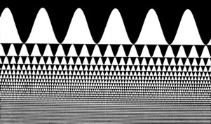
When smeared vertically this creates the sharp edged vertical bars of a square wave grating! This demonstrates the Fourier components of a square wave, with relative frequencies 1, 3, 5, 7, 9… and relative amplitudes 1/1, 1/3, 1/5, 1/7, 1/9….

A Craik-O’Brien-Cornsweet edge. The left and right regions have the same luminance but the spur-shaped luminance profile makes the right half look brighter.

Components of a Cornsweet edge are a sharp spatial step in luminance, which is very visible, plus a gradual spatial luminance ramp in the other direction, which is much less visible because the visual system is insensitive to low spatial frequencies.

How does a computer reconstitute X-ray slices to make a CAT scan? or MRI scan? Oscar Estevez (U of Amsterdam) shows this demonstration to his medical students: He rotates the cylindrical lens slowly in its own plane (around the axis of the projector beam). When the smear direction lines up with one edge of the triangle, a luminance edge appears in the smear. Smart students can guess from this that the target slide is a triangle. And a computer can guess that a target object is a brain.

Split Dots

Split Dots
With Hiro Ito

Magnified View of dot pairs. A pair of dots of different contrasts and polarities jump back and forth along orthogonal, crossing paths.

If the stimulus geometry were the only factor, all dot pairs would appear to jump along parallel paths. In fact, however, it is the vector summation of their different contrasts that make the dot pairs appear to follow different paths.

Continuous version of the split dots stimulus. All dot pairs are black & white. On the light surround at the left, they appear to drift UP to the right, and on the dark surround at the right, they appear to drift DOWN to the right.

Zebrafish

Zebrafish

with Michael Orger, Mattew Smear, and Herwig Baier (UCSF)

Zebrafish Larvae Respond to Second-Order Apparent Motion

A moving grating elicits innate optomotor behavior in zebrafish larvae; they swim in the direction of perceived motion. We took advantage of this behavior, using computer-animated displays, to determine what attributes of motion are extracted by the fish visual system. As in humans, first-order (luminance-defined or Fourier) signals dominated motion perception in fish; edges or other features had little or no effect when presented with these signals. Humans can see complex movements that lack first-order cues, an ability that is usually ascribed to higher-level processing in the visual cortex. Here we show that second-order (non-Fourier) motion displays induced optomotor behavior in zebrafish larvae, which do not have a cortex. We suggest that second-order motion is extracted early in the lower vertebrate visual pathway.

Ted Adelson’s apparent motion of a square wave minus its fundamental. Features (such as edges) move to the right but Fourier motion energy moves to the left. Result: Humans see motion to the left. So do zebrafish, responding to the Fourier motion energy, even though they have no cortex but only a primitive optic tectum.

[quicktime width=”500″ height=”300″]http://quote.ucsd.edu/anstislab/files/2012/11/zebrafish3.mov[/quicktime]

In this second order stimulus the moving bars are defined only by texture, not by luminance; on each frame, every 8th column reverses in contrast. There is no rightward motion energy, yet zebrafish will swim along to follow this stimulus.

Fourier Fun

Fourier Fun

It is well known that the shadow of the tip of a rotating rod traces out a sine wave:

SINEWAVE

It’s also well known that the Fourier components of a square wave are:
So consider a rotating arm, of length 1 and rotation rate 1.  On the arm a hand of length 1/3 rotates at a rate of 3.  On the hand a finger of length 1/5 rotates at a rate of 5… and so on.

Question:  What path in space will the tip of the set of rotating arms trace out?

SQUARE WAVE

The Fourier components of a sawtooth wave are:

SAWTOOTH WAVE

And here is a full wave rectified sinewave:

RECTIFIED SINEWAVE


Peripheral Acuity


With strict fixation of the center spot, each letter is equally legible because it is about ten times its threshold size. This is true at any viewing distance. Chart shows the increasingly coarse grain of the retinal periphery. Each letter is viewed by an equal area of visual cortex (“cortical magnification factor”) (Anstis, S.M., Vision Research 1974).  

The left hand picture shows the San Diego skyline. The right hand picture is progressively blurred from the center to the periphery. When fixated at their respective centers, both pictures look equally sharp because the progressive blurring in the right hand picture just matches the progressive loss of acuity with eccentricity caused by the increasingly coarse grain of the peripheral retina.

The retinal image undergoes barrel distortion in the retinal ganglion layer and in the visual cortex V1. This barrel distortion, or greater magnification of the center than the periphery, reflects the “cortical magnification factor”.

How Babies and Fish See Colors

What do babies and fish have in common?

Patrick Cavanagh, Stuart Anstis, Daphne Maurer, Terri Lewis

Fishes and babies can neither read nor speak, yet we persuaded them to tell us what colors they see. We showed them special moving colored patterns on a computer screen. If they saw the colors, the babies followed the movement with their eyes, and the fishes followed it by moving their whole bodies, swimming after the patterns in an innate optomotor response.

We made a movie only four frames long which looped repetitively. Each frame filled the whole computer screen with vertical colored stripes. In the left hand column, the stripes at Time 1 were light red and dark green, and at Time 2 they were light & dark yellow. The yellow stripes were shifted sideways by half a bar width, so the stripes appeared to jump sideways — but which way? The brain could not pair up succcessive stripes on the basis of color, because all the stripes at Time 2 were the same color (yellow). So the brain had to pair them up on the basis of luminance. If the red stripes were lighter than the green, the stripes appeared to jump to the right toward the nearest light yellow stripe (left-hand column). If the red stripes were darker than the green, the stripes appeared to jump to the left toward the nearest dark yellow stripe (right-hand column). The movie translates lightness into motion.

We titrated red against green luminance until, at equiluminance, perceived motion disappeared for adult obsesrvers, babies stopped making pursuit eye movements, and fishes stopped swimming in circles. Red-deficient observers rquire more red to make an equiluminous mathc, and green-deficients require more green. We were able to identify individual color-defective babies. Conclusions: Outputs from cones into the luminance pathways were in place within the first months of life. Also, guppy fish are more green-sensitive and less red-senstivie than humans.

Motion Breaks Camouflage

[quicktime width=”500″ height=”400″]http://quote.ucsd.edu/anstislab/files/2012/11/Camoflouge.mov[/quicktime]
The hidden word is well camouflaged until it moves, whereupon the visual system easily picks it out. That’s why gazelles freeze when predators are about!